
The Fabrica: When Anatomy Became Art
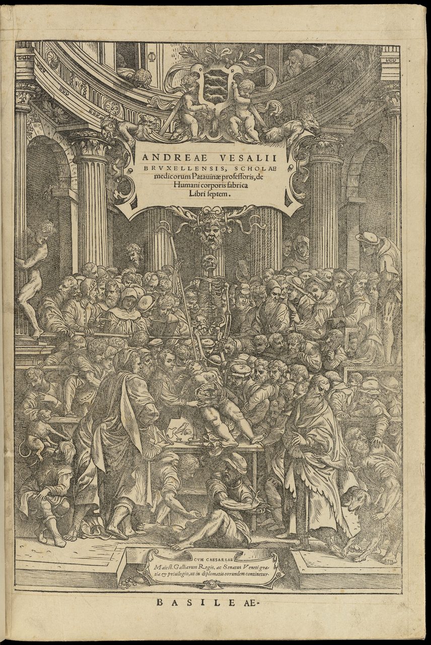
In June 1543 a printer in Basel finished a book that a professor at Padua had spent years assembling: seven books of human anatomy, illustrated with woodcuts so accomplished that people still argue about who drew them. Anatomy had been art long before 1543, so the title above is the received phrase rather than this page's claim. What made De humani corporis fabrica matter was not that it was beautiful. It was that Vesalius had cut the bodies himself, printed what he found, and put the pictures where a stranger in another country could hold them up against a corpse and disagree. He was also a Galenist who had helped bring Galen to press, who printed one of Galen's errors inside his own book on purpose, and who went on annotating his own copy for years afterwards, mostly for the Latin rather than the anatomy. This is the book, the bodies it was built from, and what it actually changed.
A book is a strange kind of instrument. It measures nothing and it has no moving parts, and yet in June 1543 a printer in Basel produced one that changed what a claim about the human body had to do before anyone was obliged to believe it. Its author was Andreas Vesalius, professor at the medical school of Padua. Its title was De humani corporis fabrica libri septem, Seven Books on the Fabric of the Human Body. What was new in it was not the looking. What was new was that the man who looked had done the cutting himself, printed pictures of what he found, and attached to those pictures an argument that a reader in another country, who had never met him and had no reason to like him, could hold up against a body and reject.
That is the version worth telling, and it is not quite the version most of us were taught. In the textbook version Vesalius arrives at Padua, discovers that Galen was wrong about everything, corrects two hundred errors and inaugurates modern anatomy. The real one is slower and stranger. He trained as a Galenist and helped bring a Galenic textbook to press. His own earlier plates carried the errors he would later reject. He printed a picture of one of Galen's mistakes inside the Fabrica on purpose, as an exhibit, and the picture outlived the argument and went on carrying the mistake into later books that copied the figure and left out what it was there to prove. He hesitated for years before he said anything at all. And the two hundred was his own boast about a single demonstration, not anybody's count of what he fixed. All of that is on the record, and none of it makes the book smaller.
01Pictures Copied Out of Other Pictures
To see what the Fabrica did, you have to know what an anatomical picture was for before it. Our own research file states the medieval position plainly: anatomical manuscripts reproduced stylized diagrams based on Galenic tradition rather than on direct observation, and the schemata it names, the frog-like squat figures, "persisted for centuries with minimal reference to actual dissection." That last phrase is the crucial one. These were not failed attempts to record a body. They were not attempts to record a body at all. They were copies of other pictures, and a copy answers to its exemplar rather than to a corpse. A reader who suspected one of being wrong had nothing to check it against except another copy.
Nor was opening a body a respectable thing for a learned man to do. By the medieval and early modern period surgery had separated from medicine and was practised by barber-surgeons, a trade whose sign, the red and white pole, is a memory of bloodletting. The physician's authority came from texts. The cutting was somebody else's job and somebody else's standing.
Susan Standring describes the oft-repeated image of a late medieval public dissection, and what it shows is a hierarchy: "the highest point in the figure is the seated lector, quoting from a text (usually based on Galen or Avicenna), physically separated from an ostensor standing below him who indicates features that the barber surgeon, incisor or sector, has revealed in the abdominal cavity of a corpse lying on a trestle table." Three men, three ranks, one body. The lector reads from the authority, the ostensor points, and the sector, the barber-surgeon, does the cutting. Her example is the woodcut in the 1493 Italian edition of de Ketham.
Then she says what the picture is actually admitting: "Most of the 'audience' show little or no interest in the proceedings, and the lector looks ahead, not at the corpse: the implication that there is little to be learned from the physical exploration happening in front of them is clear."
The man with the authority is not looking at the body.
The text those pictures answered to was Galen's, and Galen's human anatomy carried a structural problem that nobody could see from inside the tradition. Galen built his descriptions largely on animal dissection, principally Barbary macaques and pigs, because the dissection of human bodies was not practised in the Roman world of his time. He was an acute observer of what was actually in front of him. The errors entered where he assumed that what he found in an animal held in a person. Our own file makes the same point in a single line: Galen had dissected primarily animals, not humans. Every reader after him inherited those descriptions as descriptions of us.
02The Professor Who Cut
What Vesalius changed first was not how anatomy looked. It was who held the knife. Our own file's verified claim is that he established modern anatomy by "correcting Galenic errors through direct human dissection," and the operative word is direct. He cut, personally, and read the body instead of the book. The title page of the Fabrica names him Scholae medicorum Patauinae professoris, professor of the school of physicians at Padua, and by the conventions of the trade a professor with his own hands inside a cadaver was doing a barber-surgeon's work.
Susan Standring, writing in the Journal of Anatomy, describes what that produced: "As he worked, Vesalius accumulated empirical evidence of 'discrepancies' between the anatomy he saw in the human cadaver and the anatomy that he read in Galen's notes." The quotation marks around discrepancies are hers, and so is the emphasis, which in her text falls on saw and read. Of the book that came out of it she writes that it was "seven books in which he challenged the belief that Galen had described human cadaveric anatomy." That is quoted as a clause on purpose. Vesalius is not its grammatical subject, and this page will not make him one inside quotation marks, because the words between the marks have to be the words the source printed.
Seeing set against reading. That is the whole break, and everything else on this page is a consequence of it. He was twenty-eight when the book appeared, on the count Zampieri and colleagues give; some sources make him twenty-nine, counting from his birth date differently or working from a different birth year.
He did not arrive and detonate. In his Letter on the China Root of 1546, written to defend his corrections against the Galenists, Vesalius says that he lectured at Padua on Galen's On the Bones three times before he dared to call attention to Galen's mistakes.
Read that slowly. He taught Galen's account of the bones, in public, from the chair, three separate times, before he was willing to point at what was wrong with it. The best single line in the case against the heroic Vesalius is Vesalius's own, and it is a line about hesitation. It sits at Tier 2 here for a specific reason: the statement reaches this page through the secondary literature rather than from the primary text, which was not read for it. The letter itself is available in English from Cambridge University Press, translated by Daniel H. Garrison, and a reader who wants to check the sentence in context can go there.
When he did point at them, it was in public and three years before the book. Standring records that in January 1540 the students at the University of Bologna invited him to perform a public dissection, and what followed: "In front of an audience of some 200 spectators in the Church of San Francesco, he openly challenged Galen's anatomical descriptions, angering the orthodox Galenic anatomist Matthaus Curtius (Matteo Corte) who was lecturing with him."
Every part of that sentence is worth holding on to. The invitation came from the students rather than the faculty. The room was a church. And there was a specific man standing beside him, reading the text aloud while Vesalius cut, whose life's work the demonstration was contradicting. Matthaus Curtius, also written Matteo Corte, is the opponent this story usually does without, and without him the Galenists are an abstraction rather than a colleague who was angry.
Three years later the same argument arrived as a picture, on the first page any reader would see. Standring states what the Fabrica's title page does with the old arrangement: "The title-page of the Fabrica abandoned the previous conventional depiction of lector, ostensor and sector and showed Vesalius assuming all three roles as he dissected a female corpse in a crowded theatre (Fig. 5) (see Sawday, 1995)." The figure number and the citation inside that sentence are hers.
The three ranks of section 01 collapse into one man, and the collapse is the book's opening argument, made before a word of anatomy. The woodcut at the top of this page is that title page.
Two cautions, and both make the claim smaller and truer. First, the novelty is not that a professor held a knife. Standring's own aside, citing Cynthia Klestinec's study of dissection in Renaissance Venice, is that combining the roles of lecturer and dissector was already a common practice in public demonstrations. What was new was putting the collapse of the three roles into a printed picture and making it the first thing the book says. Second, the fuller reading of what else that woodcut is arguing belongs to Jonathan Sawday's The Body Emblazoned, which is where Standring sends her reader and which this page has not read. It is listed below as where that argument is set out, and nothing here is attributed to it.
03Where the Bodies Came From
A book built on direct human dissection needs a supply of human bodies, and the sourced account of where Vesalius got his is quieter, and worse, than the grave-robbing legend. Standring states it in one sentence: "Vesalius had access to a relatively plentiful supply of cadavers, possibly supplemented by grave robbing, but mainly reflecting his good working relationship with Marcantonio Contarini, a sympathetic judge in the criminal court who apparently arranged the time of executions according to the anatomist's needs."
Two things in that sentence are true at once and both belong in any honest account. The first is that this was official cooperation with a court and not a crime: the arrangement ran through a judge of the criminal court rather than around him. The second is what the arrangement means once you stop and read it. A criminal court was scheduling the deaths of human beings to suit a professor's teaching calendar. Standring's hedge, apparently, is hers, and it is kept here, because nobody has the minutes of that understanding.
Vesalius had been a student in Paris before he was a professor in Padua, and Standring records that too, in a single sentence that also names a second city: "Vesalius was probably describing the behaviour of many similarly enthusiastic anatomy students when he recalled picking over corpses in the charnel house by the public gallows at Montfaucon and at the Cemetery of Innocents while he was a student in Paris, and disarticulating a dried skeleton found by the roadside when he was a student in Louvain (Saunders & O'Malley, 1950)." Montfaucon was the great gibbet of Paris. Note her framing, which is harder on the period than singling him out would be. This was ordinary student behaviour.
What that kind of legal cooperation was made of is easiest to see somewhere other than Padua, because that is where the documented example sits. Standring's is English. An Act of Parliament in 1540 joined the Guild or Fellowship of Surgeons and the Barbers' Company into the Company of Barbers and Surgeons of London, an occasion Holbein painted with Henry VIII handing the charter to Thomas Vicary, and the new company was permitted to dissect four bodies a year at Barber Surgeons' Hall in Monkwell Street, close to Newgate Prison. Where those four bodies came from was written into the sentence of the court: "Convicted murderers could be dissected by anatomists after they were hanged: 'opening of the body' was a fate reminiscent of the medieval practice of hanging, drawing and quartering of traitors and was an additional punishment inflicted upon the corpse."
Dissection there was not a loophole in the death penalty. It was more of the death penalty, carried out after death.
That is Tudor London, and this page does not carry it to Padua. Vesalius's cadavers came through a criminal court of the Venetian Republic, and Jacob Karrer, further down this page, was beheaded at Basel rather than hanged in London. What holds generally is that in sixteenth-century Europe dissection after execution was a legally sanctioned addition to the penalty; the specific provision quoted above is England's, and it is not the law Vesalius worked under.
Two things about the bodies stay at credible rather than verified, and in both cases the qualifier is the claim rather than a softening of it.
The first is grave robbing. Standring's phrase is "possibly supplemented by grave robbing." No source consulted for this page establishes that Vesalius personally robbed a grave in Padua. What can be said is that the supply may have been supplemented that way. What cannot be said is the sentence everybody wants, which is that Vesalius was a body snatcher.
The second is the gibbet story, which is genuinely his and genuinely unsettled. As a student he took the remains of an executed criminal from a gibbet outside a city wall, returned after dark to carry off the trunk, and built a complete skeleton from the bones, with the help of his friend the cosmographer Gemma Frisius. The episode descends from Vesalius's own account. Where it happened is where the sources part company: some place it at the gallows hill outside Louvain, others at Montfaucon in Paris.
Standring narrows that without closing it, and the narrowing is in the sentence quoted above. She puts the charnel-house scavenging at Paris and a separate student episode at Louvain, so a peer-reviewed source does place bone-taking at Louvain. But what she describes there is a dried skeleton found by the roadside, with no gibbet, no return after dark and no Gemma Frisius. The two accounts are compatible and may well be one episode told twice, and she does not say so, so this page will not say it either. The standard biography, C. D. O'Malley's Andreas Vesalius of Brussels, 1514-1564, was not read for this page. The disagreement between the secondary accounts is real rather than careless.
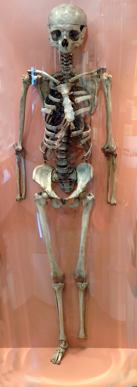
One of those bodies has a name, and you can still go and look at him. In 1543, while Vesalius was in Basel seeing the Fabrica through the press, a man was beheaded in the city, and Vesalius articulated a skeleton from his bones. It is still on display at the Anatomical Museum of the University of Basel. Zampieri and colleagues put it this way: "The skeleton he constructed from the bones of an executed criminal, Jacob Karrar, is still preserved in the Anatomy Museum of Basle's University."
The name is spelled three ways across the sources, Karrar in that paper, Karrer on the museum photograph's own record, Jakob Karrer von Gebweiler elsewhere. They are one man, and this page prints Karrer. The variance is in the paperwork, not in what happened to him. He is the point at which the phrase "his cadavers came from executed criminals" stops being a sentence about supply and becomes a sentence about a person, which is the correct discomfort to feel and the reason he is named here.
04Basel, June 1543
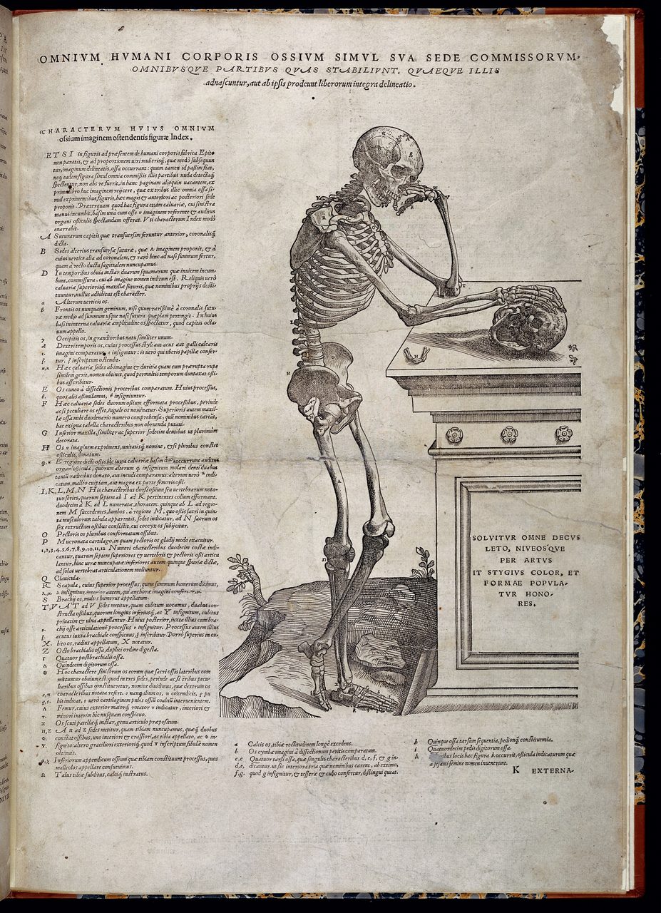
The book is a specific object with a specific date on it. Its full title is De humani corporis fabrica libri septem, Seven Books on the Fabric of the Human Body, and it was printed at Basel by Johannes Oporinus. The imprint the US National Library of Medicine records for its plates gives the month: "Basileae : Ex officina Joannis Oporini, anno salutis reparatae 1543. Mense Iunio." Basel, from the workshop of Johannes Oporinus, in the month of June 1543. The Getty Research Institute reads the colophon of its own copy the same way, which is a second institution and a second physical book saying June.
The printer's name carries a small pleasure, and the record that carries it carries an error. The Rijksmuseum describes its sheet as "Titelprent voor: Vesalius, Andreas. De Humani Corporis Fabrica. Bazel: Jan Herpst, 1543." That sheet is not the Fabrica's title page. Its own cartouche ends EPITOME, so what the museum is describing is the title page of the companion volume. Institutional records are the best evidence available here and they are not oracles, which is a refrain on this page rather than a complaint. What the museum has exactly right is the printer. Jan Herpst is not a mistake: Oporinus was born Johannes Herbster, and Oporinus is the Greek form of Herbst, so the museum is recording the man rather than the imprint.
Seven books, as the title says. Book I is the bones and Book II the muscles. Our own file does not enumerate the remaining five and this page will not invent them. In the same year Vesalius issued a companion volume, the Epitome, De humani corporis fabrica librorum Epitome, a condensed version of the seven-book work, also out of Basel.
Two figures people reach for about this book have to be handled differently from each other, because one of them is open and the other has actually been counted.
How many illustrations the Fabrica contains is not settled, and the disagreement has a shape worth seeing. Zampieri and colleagues write that there are more than 250. Standring, counting for a different purpose, says the seven books used over 200 illustrations of Vesalius's own dissections. The Getty Research Institute's record for its own copy of the 1543 edition gives a bibliographer's figure instead: 204 illustrations in the text, with the illustrated title page listed separately from them. Other reputable secondary sources say more than 200, and others over 300. No authoritative single count was found for this page, and the likeliest reason the figures disagree is that the counters are counting different things, a bibliographer working through one physical copy and listing the title page apart, a historian counting plates. So the honest forms are the attribution or the floor: more than 250 on Zampieri and colleagues' count, or more than two hundred as a floor, with the tally still open. A precise total stated flatly would be borrowing confidence from somewhere it does not exist, and 204 is one library's count of one copy rather than the answer.
How many copies survive does have an answer, because a team went and looked. A worldwide census of the Fabrica by Dániel Margócsy, Mark Somos and Stephen N. Joffe was published by Brill in 2018. The project's own page at Cambridge says the team documented "over seven hundred copies of these two editions around the world," meaning the 1543 and the 1555, recording bibliographic information, ownership records and records of handwritten annotations, and examining "how readers studied, annotated and critiqued the book from 1543 to 2017." A per-edition split of that total appears in secondary reporting of the census; it was not confirmed against the census itself and it is not printed here.
05The Corrections, and Two You Can Check on Your Own Body
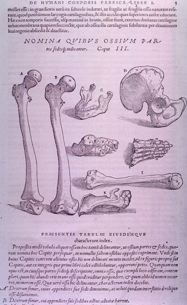
The corrections that matter most for an article about evidence are the ones a reader can test, and Zampieri and colleagues set four of them out in a single sentence with the reason attached: "That Galen made many mistakes was a foregone conclusion, since human and ape anatomies are very different. Between Galen's errors, Vesalius showed that the sternum consisted of three sections, instead of seven, that the mandible consisted of one bone, instead of two, that the 'rete mirabile' did not exist in man, and that nerves were not hollow."
Two of those four are settled by your own hand. Put a hand flat on your breastbone and you are touching the first. Run a finger along your jaw from the ear to the chin and you are touching the second. What makes the pair so useful is that neither is a careless error. Both Galenic figures are what you find in an animal, which is Zampieri and colleagues' own reason, stated in the first half of that sentence. The mechanism of the entire Galenic problem is visible in those two bones: a correct observation of a macaque or a pig, carried across to a human being on the assumption that the two would agree.
The third item in that sentence is the rete mirabile, described by Zampieri and colleagues as "a network of vessels at the base of brain that Galen supposed existed in humans, while in fact it exists only in other mammals."
Galen had not merely described a structure that is absent. He had built physiological theory on it: the structure was load-bearing for the whole Galenic account of the spirits. Vesalius's denial of it in people is among his most consequential single corrections for exactly that reason. Remove the structure and the theory has nowhere left to happen. This is also the correction that will come back later on this page and complicate everything, because of what Vesalius chose to print alongside it.
The fourth item, that nerves were not hollow, is given by Zampieri and colleagues in exactly those words and no further. No source consulted for this page elaborates on it, so it is recorded here and left at that.
One more correction has a date on it, and the date is the whole point. Galen held that blood passed from the right ventricle to the left through pores in the wall between them. In 1543 Vesalius did not deny those pores. Zampieri and colleagues introduce his first-edition sentence by saying that he did not deny the patency of the interventricular septum but seemed, at least, doubtful, and then quote him: "Thus we are compelled to astonishment at the industry of the Creator who causes the blood to sweat from the right ventricle into the left through passages which escapes our sight." That is a man declining to contradict Galen, and admiring the workmanship of a passage he cannot find.
Twelve years later he contradicted it, and the sentence is more careful than its reputation. Zampieri and colleagues quote the second edition: "However much the pits may be apparent, yet none, as far as can be comprehended by sense, passes through the septum of the heart from the right ventricle into the left [...] although they are mentioned by professors of anatomy since they are convinced that blood is carried from the right ventricle to the left. As a result, as I shall declare more openly elsewhere, I am in no little doubt regarding the function of the heart in this part."
The bracketed ellipsis there is the source's own and it is the only one in the passage. Two clauses in that sentence usually go missing, and both of them cut against the tidy version. As far as can be comprehended by sense limits the denial to what the senses can reach, rather than declaring the pores impossible. And the sentence ends not in triumph but in doubt about what the heart is doing in that place. Zampieri and colleagues say so themselves in their next line: that last part is significant, because it expresses the idea that a new physiology of the heart was emerging.
That twelve-year gap between the doubt and the denial is not a footnote. It is the clearest single measurement anyone has of how the break with Galen actually went, and it is the reason the fourth row of the table below reads differently from the first three.
| The Structure | The Galenic Account | What Vesalius Reported | How Settled It Was In 1543 |
|---|---|---|---|
| The sternum | Seven sections | Three sections | Stated in the first edition |
| The mandible, the lower jaw | Two bones | One bone | Stated in the first edition |
| The rete mirabile, a network of vessels at the base of the brain | Present in people, and load-bearing for the whole Galenic account of the spirits | Present only in other mammals, absent in people | Denied in the first edition, which also printed a figure of the Galenic human version as an exhibit of the error |
| The wall between the ventricles of the heart | Blood passes from the right ventricle to the left through pores in it | Doubt in 1543; in 1555 the pores denied, but only as far as can be comprehended by sense, and with his doubt about the heart's function in that place left standing | Left open. The denial quoted above is from 1555, not 1543 |
06The Complication: He Was a Galenist
Before the Fabrica, Vesalius worked on Galen and inside the Galenic tradition. Vivian Nutton's 2017 Routledge edition of the text he helped produce is titled Principles of Anatomy according to the Opinion of Galen by Johann Guinter and Andreas Vesalius, and it carries a chapter headed "Vesalius' preface." The young Vesalius had revised his teacher Johann Guinter's Galenic anatomy textbook. The man remembered for overturning Galen had first helped put Galen into print.
That is not a gotcha. It is the correct frame: he knew the text well enough to know where it failed.
His own earlier plates failed in the same direction, and that can be said plainly rather than hedged, because Standring states it in one sentence: "The anatomy illustrated in the Tabulae is quite clearly Galenic anatomy: for example, the chart of the arterial system included the rete mirabile and the liver was five-lobed." The Tabulae Anatomicae Sex of 1538 were six very large woodcut sheets issued as handouts for his own students, five years before the Fabrica, and they carry two of the structures the Fabrica would deny. Standring puts it more bluntly still elsewhere: Vesalius "initially hedged his bets, retaining the rete mirabile" in those sheets. This was a drift, not a conversion.
One detail in the 1538 sheets belongs to this page's argument rather than to its list of errors. Three of the six showed the portal, caval and arterial systems, from charts Vesalius had drawn while teaching. The other three showed a skeleton in life-like poses from three sides, and Standring records that they were drawn by Jan Stefan van Calcar "from a human skeleton that had been articulated by Vesalius." Five years before the Fabrica, the artist was already working from a body the anatomist had prepared with his own hands, rather than from another picture. That is the whole thesis of this page, documented in 1538. (Standring spells the artist Jan Stefan van Calcar; the Library of Congress, the Biodiversity Heritage Library and Zampieri and colleagues all print Jan Stephan van Calcar. One man, two spellings.)
That sentence is the origin of the most repeated fact about Vesalius, and it does not say what the repetition says.
The textbook version, carried across popular accounts and, it has to be said, by our own research file, is that Vesalius corrected two hundred of Galen's errors. X_5_02's summary states that he "corrected over 200 of Galen's anatomical errors," and the planning notes for this article repeated it, so the correction being made here lands on us first and on the textbooks second. What the scholarship actually reports is Vesalius's polemic: that Galen, in the course of just one anatomical demonstration, gets wrong more than 200 times in the right description of the parts, the harmony, the use and the function of human body. That trailing clause is the part that falls off in retelling, and it is the part that decides the meaning. With it, the boast is about how wrong Galen's whole account is across one demonstration. Without it, it reads as a tally of two hundred separate mistakes, which is exactly what it has become. It is a rhetorical claim about one demonstration, made by an interested party in the register of a man picking a fight in public, and it is not a modern count of the corrections in the Fabrica. No source consulted for this page counts them and arrives at two hundred, and none was found that counts them at all.
Three different two hundreds attach themselves to Vesalius in the sources used here, and they are three different quantities. Standring counts over two hundred illustrations in the seven books. Standring also puts some two hundred spectators in the church at Bologna in January 1540. Vesalius claims more than two hundred errors in one of Galen's demonstrations. Pictures, people, mistakes: the round number is the only thing the three have in common, and how easily two of them could be fused into a fourth number that nobody ever asserted is the whole subject of this section.
The round number survived and the sentence around it did not, which is a small working demonstration of the thing the rest of this article is about. A compact claim travels. Its qualifications stay behind.
And then there is the picture he printed of the mistake.
The Fabrica contains a figure of the human rete mirabile, the structure Vesalius said was not there. He printed it alongside a schematic of the real rete mirabile in hoofed animals, and he printed it deliberately: it was an exhibit of an error, the error itself set out so that a reader could see where Galen's overreliance on animals had led. Douglas Lanska has traced what happened next. The intent did not survive transmission. Later printed books copied Vesalius's figure and left out his argument, and the myth of a human rete mirabile went on living inside the illustration of the book that had killed it.
This is the sharpest thing in the whole story, and it is here precisely because it cuts against the argument this page is making. A printed picture is a powerful instrument of evidence. It is also a powerful vector for error, for the same reason: the picture travels further than the caption.
He never stopped working on the book, though not in the way the phrase usually implies. Vivian Nutton's 2012 paper in Medical History reports a find, and its abstract states it plainly: "A second edition was published in Basle in 1555, but little is known of Vesalius' activities after that date. This article discusses a recent find: Vesalius' own copy of the 1555 edition, heavily annotated in preparation for a never published third edition."
The same abstract is careful about what those annotations are, and the care has to be carried with the find. Vesalius made hundreds of changes, "the great majority being stylistic, altering the Latin words but not the overall meaning." There are also changes to the plates "to give greater clarity or to correct mistakes by the original block-cutter." And then the sentence that decides how this page may use the discovery: "There is little new anatomical material, although Vesalius continued to meditate about what he had earlier discovered."
So the annotated copy is not evidence of a man still finding new anatomy. It is evidence of something that suits this page better anyway, and Nutton says it outright in his closing line: the number of annotations shows "Vesalius' passionate concern not only for accuracy but also for the most effective way of proclaiming his new anatomical message." A man going back through his own book line by line, mostly to make it read better and print better, is a man working on how a claim travels.
The block-cutter is worth a moment. Some of what he was fixing was not anatomy at all but damage done between the drawing and the printed sheet by the man who cut the blocks. The picture and the argument can come apart in production, which is the same failure this page meets twice more: in the later books that copied his rete mirabile figure and dropped what it was for, and in a round number that outlived its own sentence.
There is a scholarly position that goes further than gradualism, and it deserves stating at strength rather than waving at. On the revisionist reading, the great Renaissance anatomists were resurrecting the anatomical projects of the ancients rather than rejecting them, choosing their subjects and their aims in a spirit of emulation. Andrew Cunningham's The Anatomical Renaissance: The Resurrection of the Anatomical Projects of the Ancients, published in 1997, is the standard statement of it. Vivian Nutton's Andreas Vesalius and his Fabrica, 1537-1564: Changing the World of Anatomy, published by Springer in 2024, is the current full-length scholarly treatment of the book itself.
The reading has a harder second half. Many of Vesalius's own contemporary readers, possibly most, took his message as a continuation of the empirical and experimental method Galen himself had advocated and could not fully practise, rather than as its overthrow. On that view Vesalius is a kind of neo-Galenist: a man who epitomised Galen's own practice of anatomy as an observational science while correcting the results Galen got with it. This is an interpretive position in the historiography and not a settled fact about what every sixteenth-century reader thought, and it is carried here as a position. It is also the reading that makes the most sense of a man who revised his teacher's Galen, lectured three times on Galen's bones before objecting, and printed one of Galen's errors as a teaching exhibit.
Put the pieces together and the shape of the break changes. Doubt about the pores in 1543. A denial in 1555 that keeps its qualifier and ends in doubt. Then hundreds of annotations for an edition that never appeared, most of them about how the Latin reads. The correction of Galen was a process, and it outlasted the famous book by more than a decade. A revolution that takes twelve years to harden one doubt into a hedged denial, and whose author then spends years on how best to say it, is a different kind of event from the one in the textbooks. It is not a smaller one. It is an event with a duration, made by somebody who could be wrong and who cared how he would be read.
07Why It Had to Be Printed
There is a control experiment for the claim that the Fabrica changed anatomy, and nobody designed it.
Leonardo da Vinci, who died in 1519, produced over 750 anatomical drawings from his own dissections, covering the cardiovascular system, the musculature, fetal development and the skeleton. They are decades earlier than the Fabrica, and by any ordinary measure of accuracy they are extraordinary. Our own file records the complication: historians debate their significance for the history of anatomy, as opposed to the history of art, precisely because they were not published or widely circulated, remaining unpublished until the eighteenth and nineteenth centuries. The file's verdict is blunt. "Leonardo's work did not directly influence the development of anatomy as a discipline; Vesalius, not Leonardo, drove the anatomical revolution that transformed medicine."
That is this page's whole argument in one comparison. Better drawings, made earlier, from real dissection, changed nothing at all, because nobody could see them.
What Vesalius had that Leonardo did not was a press, and a specific idea about what to do with one.
Sachiko Kusukawa's Picturing the Book of Nature: Image, Text, and Argument in Sixteenth-Century Human Anatomy and Medical Botany, published by the University of Chicago Press in 2012, is the scholarly statement of it. Her argument is that Leonhart Fuchs and Andreas Vesalius deployed images in order to make specific arguments about nature, producing a new kind of visual argument, and that in doing so they ran counter to the prevailing practice of copying pictures out of other books. Set that against the frog-like squat figures of section 01 and the change is exact. A picture stopped being a mnemonic attached to a text and became a piece of evidence with a claim attached, issued in enough identical copies that a stranger could disagree with it in public.
It is worth being precise about what this page's own title does and does not assert. Anatomy had been art before 1543, and Leonardo is the proof of it. What 1543 did was make anatomical art public, identical and checkable, which is a different thing and a larger one.
None of it happened in isolation, either. Ambroise Paré, working in France in the same generation, abandoned the cauterisation of gunshot wounds in favour of gentle wound treatment and tied off arteries instead of burning amputation stumps, on the strength of what he watched happen to patients. The year 1543 belongs to a wider turn toward looking, not to a lightning strike in an empty field.
08Who Drew It, and What the Landscape Is Doing
Who actually drew the Fabrica is not settled, and the way it is unsettled is instructive.
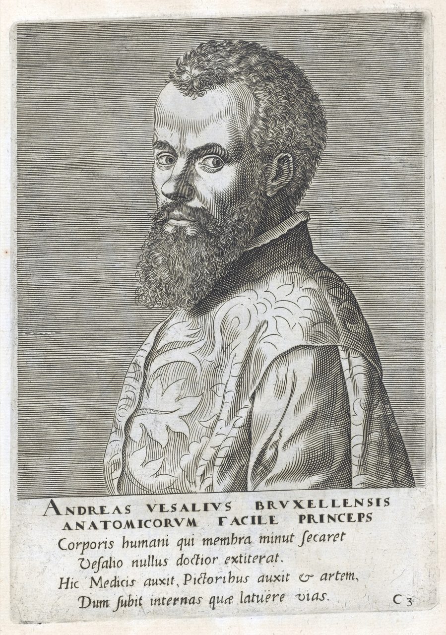
The traditional attribution is to Jan Stephan van Calcar, a student of Titian. Our own file rates it Tier 2 and says why: it rests on "stylistic analysis and circumstantial evidence," Vesalius having been in Padua where Calcar studied under Titian, with the artistic quality suggesting a trained artist. The file then states the objection in the same breath: "however, scholars argue multiple artists contributed, and the attribution remains debated."
The holding institutions disagree with each other in their own catalogues, which is worth showing rather than smoothing. The Biodiversity Heritage Library's record for the 1543 book lists van Calcar as a co-author alongside Vesalius. The Getty Research Institute's record for its own 1543 copy assigns the illustrations to him flatly, in a bibliographer's note. The Library of Congress catalogues the title-page woodcut as by "Calcar, Jan Stephan van, 1499-1546?, engraver," question mark and all. Standring says Calcar drew the skeleton figures specifically. Zampieri and colleagues say only that these pieces of art have been attributed to Jan van Calcar. And the Boston Public Library's record for its copy of the 1555 edition lists both Titian and Calcar as woodcutters while marking each of them, in the metadata itself, attributed name.
Six statements, six degrees of confidence, one book, and the most careful of the six is a cataloguer who wrote the word attributed into the record. Our own file's closing line is the one this page will stand behind: whoever the artist or artists, the illustrations demonstrate the Renaissance synthesis of empirical observation and artistic mastery.
There is a compositional fact about the muscle plates that changes how you look at them.
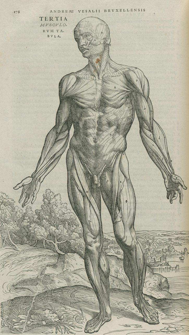
The muscle figures at the beginning of Book II are fourteen full-page plates showing a single body in progressive stages of dissection, and their landscape backgrounds are not fourteen separate scenes. They join edge to edge into a continuous panorama. Two dedicated papers in Medical History address it: G. S. T. Cavanagh's "A new view of the Vesalian landscape" of 1983, and Martin Kemp's "A drawing for the Fabrica; and some thoughts upon the Vesalius muscle-men" of 1970.
Three hedges hold and should be visible. The terrain is commonly identified as the countryside west of Padua, often the Euganean hills, but that identification is an inference from the topography rather than a documented statement by anyone in 1543. The figures reverse and reorder relative to the frieze, which is why the join is not obvious to anyone turning the pages of a bound copy. And the original recognition of the panorama is credited in the secondary literature to a scholar this page could not tie to a checkable identifier, so no name is attached to it here.
Whatever the intention behind it, the effect is that fourteen sheets read as one continuous act of dissection rather than as fourteen separate studio poses.
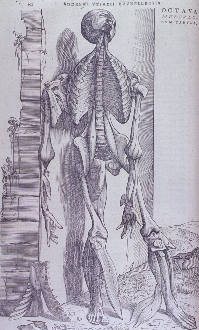
That the figures stand in a landscape at all is confirmed from outside the scholarship, in the flattest possible language, by the catalogue of the US National Library of Medicine. Its abstract for the third muscle tabula reads, in full: "Standing nude male figure in landscape; muscles delineated." Its abstract for the eighth reads: "Full length, front view, body leaning against stone wall. Muscles of face, neck, arms and legs exposed. Chest cavity open revealing rib cage and vertebrae." The records list both sheets as woodcuts, black and white, at 42 by 28 and 41 by 27 centimetres, printed at Basel by Oporinus in June 1543, and both name Vesalius as a contributor while the record for the third also names Calcar.
A cataloguer describing what is on a sheet is not interpreting anything, which is exactly what makes the record useful. The landscape is not a scholar's reading imposed on the plates four centuries later. It is what is printed on the paper, and a librarian wrote it down without being asked to have an opinion about it.
09The Theatre and the Blocks
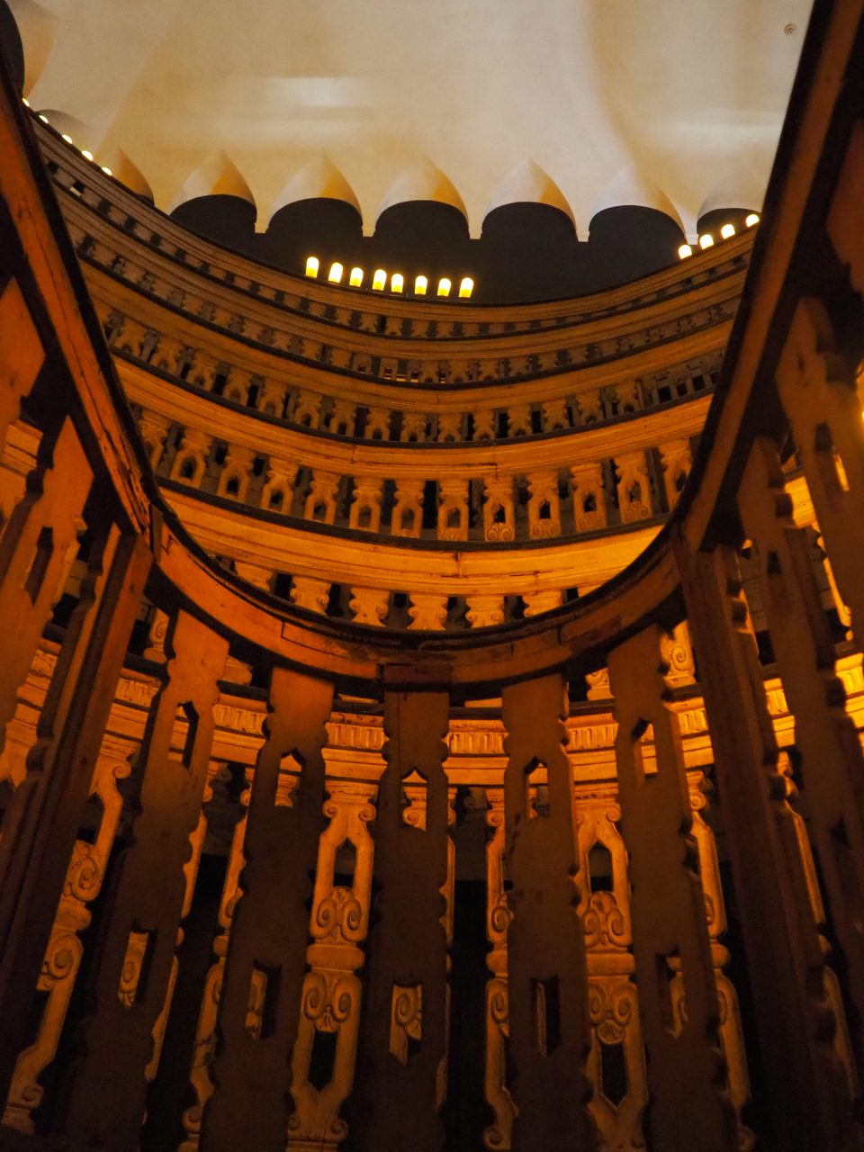
Dissection did not stay a private act performed on a table with a crowd pressing round it. The first permanent anatomical theatre was built at the University of Padua in 1594, designed by Girolamo Fabricius, and purpose-built dissection theatres went on to become centres of public education and spectacle, with demonstrations attended by students, physicians and the public.
The date matters more than it looks. 1594 is fifty-one years after the Fabrica and thirty years after Vesalius died. He never taught in that room, and no photograph of it is a photograph of anything he did. What the theatre records is what his book's audience eventually got built for them: a piece of architecture whose entire purpose was to let a large number of people watch one body being opened.
The blocks themselves had a long life and a bad end. The original Fabrica woodblocks survived for four centuries, passing from Basel to Munich, and were used one last time in 1934 to print the Icones Anatomicae, produced by the Bremer Press for the New York Academy of Medicine and the University of Munich. The blocks, and the German edition of the Icones, were destroyed in a 1944 bombing raid on Munich.
The accounts consulted for this page give different dates within that year, and this page does not adjudicate between them. The year is what can be said. Four centuries of survival, one raid, and the objects that made the argument possible are gone. What remains is what was printed from them, which is the point this article has been making about printing, delivered by accident and at a price nobody would have chosen.
10What Came After the Woodcut
The next real change in anatomical illustration was not a change of style. It was a change of material, and it quietly solved the problem this article keeps circling.
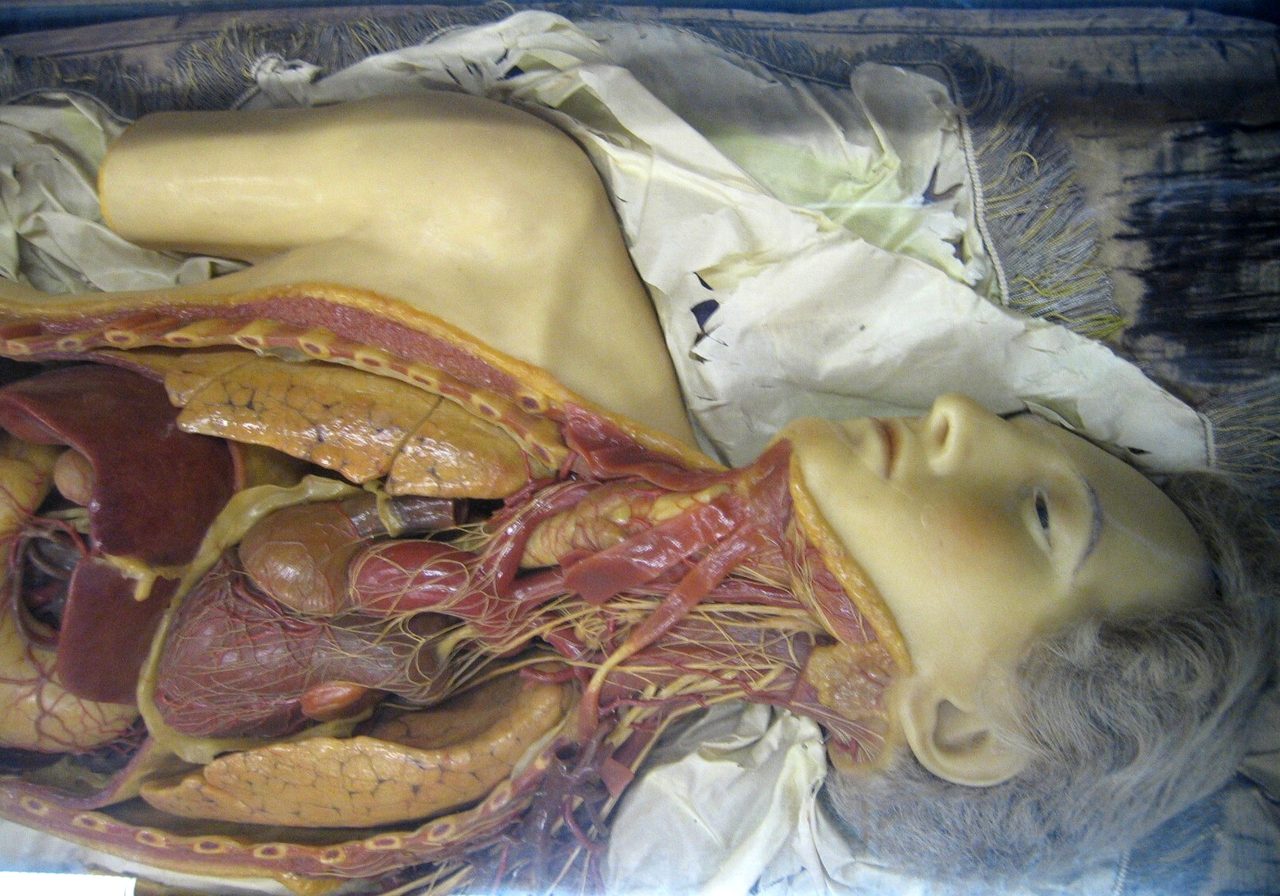
In eighteenth-century Florence, at La Specola, Felice Fontana and Clemente Susini created thousands of detailed wax anatomical models for teaching, and their significance is not aesthetic. Wax removed the dependence on cadaver availability. For the first time a teaching anatomy could exist that did not require a body this week, which makes La Specola a genuine turning point in the provenance thread rather than a curiosity.
The wax tradition also produced the Anatomical Venuses, reclining female figures that could be opened to reveal the internal organs. Our own file's reading of them is worth quoting as the judgement it is: they "combined scientific instruction with idealized female beauty in ways that reflected and reinforced gendered views of the body," and "the idealized Anatomical Venuses encoded gender norms." That is our file's assessment rather than an established finding, and it is offered here as such.
Three centuries after the Fabrica, the English-speaking world got its own standard anatomy, and repeated the argument about the pictures almost exactly.
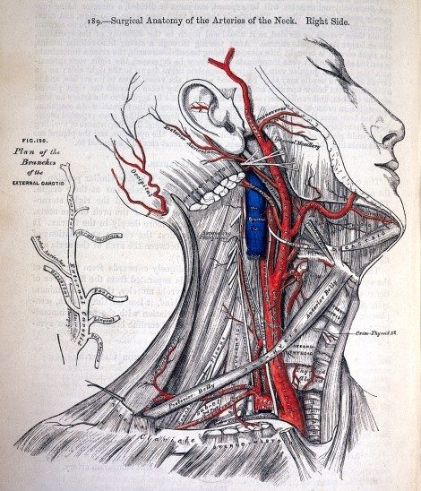
Henry Gray's Anatomy, Descriptive and Surgical appeared in 1858, illustrated by Henry Vandyke Carter, and became the standard English-language anatomy textbook; it is universally known now as Gray's Anatomy. The Wellcome Collection's record for a first-edition plate names Henry Vandyke Carter in its Artist field, which is a small institutional acknowledgement of the illustrator and, as it turns out, a pointed one.
Gray and Carter fell out over exactly that. When Gray received the proof of the title page, which set his name as author and Carter's below it as illustrator in the same size type, he marked it up demanding that Carter's name be set smaller and stripping Carter of his titles. The marked-up proof survives at the Royal College of Surgeons. Ruth Richardson's The Making of Mr Gray's Anatomy, published by Oxford University Press in 2008, is the study of the episode, and her published position is that the originality of the first edition resided in the illustrations.
That is the Calcar question again, three hundred years later, with the evidence attached. When a book's authority is carried by its pictures, whose book is it? In 1543 we cannot answer, because the record is thin and the institutions disagree. In 1858 we can, because the argument survives in pencil on a proof sheet.
The profession that argument implies arrived in the twentieth century. Max Brödel, 1870 to 1941, came to Johns Hopkins from Leipzig in 1894 and in 1911 founded the Department of Art as Applied to Medicine there, the first academic department of medical illustration in the world, endowed by Henry Walters. Our own file's verified claim is that his techniques, including carbon dust rendering and an anatomical accuracy obtained by directly observing surgery and dissection, established the professional standards that define the field, and that the Association of Medical Illustrators, founded in 1945, traces its intellectual lineage to him.
Frank Netter, 1906 to 1991, whom people called the Michelangelo of medicine, produced over 4,000 medical illustrations characterised by clarity, and his atlases remain widely used. The epithet is what he was called rather than a fact about him. His work is in copyright, which is why none of it appears on this page.
11The Thread Nobody Chose
Follow the bodies forward and the story stops being about one book.
Standring records what happened to the supply Vesalius had worked from. By the middle of the seventeenth century, she writes, "The supply of bodies from the gallows could not satisfy the demands for corpses for education and research." The date is hers too, from the sentence before that one. Demand grew, the legal source did not, and the gap between the two is the whole subsequent history of where anatomy schools got their material.
That history has a literature of its own and it is not this article's, but it should be named rather than gestured at. Ruth Richardson's Death, Dissection and the Destitute, first published by Routledge and Kegan Paul in 1987 and reissued by the University of Chicago Press in 2001 with a new afterword, is the standard study of Britain and the Anatomy Act of 1832. Michael Sappol's A Traffic of Dead Bodies: Anatomy and Embodied Social Identity in Nineteenth-Century America, published by Princeton University Press, is the standard study of the American side. Both were consulted for this page as bibliographic records rather than read, so no figure, date or episode on this page is attributed to their contents.
One scope note, in the open, because this is exactly the kind of material that gets transplanted. Our own research file's counter-arguments include the sentence that the use of executed criminals' and enslaved persons' bodies for dissection reflects power relations. That is a claim about the history of anatomy as a whole. It is not a claim about Vesalius, about Padua, or about 1543, and no source consulted for this page places enslaved people among Vesalius's dissection subjects. The scholarship that genuinely covers enslaved and destitute bodies, Richardson on Britain and Sappol on nineteenth-century America, concerns other centuries and another continent. It is named above, in its own place, and it is not carried backward onto 1543.
The worst case in the history of anatomical illustration is documented, twentieth-century, and still on the shelf.
Eduard Pernkopf's Topographische Anatomie des Menschen, published in English as the Atlas of Topographical and Applied Human Anatomy, was prepared over about twenty years at the University of Vienna by Pernkopf and a team of artists, and is still regarded by some surgeons as anatomically unsurpassed. Investigations in the 1980s and 1990s established that Pernkopf was an active National Socialist functionary at the university, and that among the bodies depicted in the atlas were those of Nazi victims. No count of them appears here; none was verified for this page, and a number invented for effect would be its own small desecration.
The response has not been the obvious one, and both positions are held by serious people.
Sabine Hildebrandt's 2006 paper in Clinical Anatomy carries its conclusion in its own title: "How the Pernkopf controversy facilitated a historical and ethical analysis of the anatomical sciences in Austria and Germany: A recommendation for the continued use of the Pernkopf atlas." Against destruction or withdrawal, the argument is that the images exist, that they are anatomically valuable, and that the honest course is to use them with the history stated. In 2019 a surgical team facing a specific operative problem published a case study in Surgery describing how they consulted historians of Nazi medicine, bioethicists and experts in Jewish law before deciding whether to consult the atlas at all.
This page does not pick a side. A reader who believes the atlas should never be opened again, and a reader who believes that refusing to use it wastes the only thing the victims' bodies can still be made to give, are both reasoning from real principles, and neither has been shown to be wrong.
The Visible Human Project, run by the US National Library of Medicine from 1994, produced complete cryosectioned datasets of a male and a female cadaver, the male body sectioned at one-millimetre intervals. That body was Joseph Paul Jernigan's. He was executed by lethal injection in Texas on 5 August 1993, having agreed to donate his body at the prompting of a prison chaplain and without knowing about the project. The female dataset came from an anonymous donor who died of cardiovascular disease. Catherine Waldby's The Visible Human Project, published by Routledge in 2003, is the book-length scholarly treatment of the ethics.
The line this page will draw is structural. Four and a half centuries after a judge in Padua timed executions to an anatomist's calendar, the flagship digital anatomy of the twentieth century again rests on the body of an executed man whose consent is arguable. The line it will not draw is that this was anyone's intention, that there is a lineage running from 1543 Padua through Vienna to a Texas execution chamber, or that any of it is Vesalius's fault. No source on this page asserts such a lineage and none exists on the record. What exists is a pattern, which is worse, because a pattern requires nobody to plan it.
Our own file states the general case in a sentence this article has spent its whole length illustrating: "The history of anatomical art reveals that visual representation is never neutral. Choices about what to depict, how to pose bodies, which bodies to use as models, and what to emphasize or omit reflect the social values and biases of their creators." It closes by noting that modern medical illustration still faces the same questions about representation, in skin colour, body type, gender and age diversity across anatomical atlases.
The muscle men posed in their landscape, the Anatomical Venuses, the Pernkopf plates and a diversity audit of a current atlas are the same question asked in four different centuries. Who is being depicted, by whom, and what has been left out of the frame.
12What a Drawing Does That a Photograph Cannot
Which leaves the question of whether any of this still matters, now that a body can be photographed, scanned and rendered in three dimensions.
Our own file's closing judgement grants the transformation and then turns on it. Three-dimensional visualisation, virtual dissection, surgical simulation and augmented reality are transforming the field, "however, hand-drawn medical illustration remains valued for its ability to selectively emphasize clinically relevant structures in ways photography and 3D imaging cannot."
That is the argument of 1543 with different equipment. A photograph records everything in front of it and therefore argues nothing. A drawing chooses what to show, and choosing is what turns a picture into a claim. It is also what makes a picture capable of being wrong, which is the price of the whole method, and Vesalius's rete mirabile is the receipt.
One caveat belongs here, and this page's own illustrations supply it. The case for print is that it makes enough identical copies for a stranger to hold one up against a body. Identical is doing a great deal of work in that sentence. The Gray's figure reproduced further up is coloured one way in the copy shown here, red arteries and a blue jugular, and another way in a second digitised copy of the same 1858 first edition, where figure 190 is pink and the jugular is not coloured at all. Two copies of one edition, not identical. The printed page is a far better instrument than a hand-copied one, which is the whole of the claim this page has been making, and it is not the flawless one the claim is sometimes stretched into.
The people who do the choosing occupy a precarious professional position. Trained medical illustrators, typically holding master's degrees from accredited programmes, provide expertise that stock photography and generated imagery cannot replicate, while economic pressure and the proliferation of free digital images threaten the viability of the profession Brödel built.
Generative systems have begun producing medical illustrations, and what happens next is genuinely open. Whether such a system can match the selective emphasis, clinical accuracy and pedagogical clarity of a trained medical illustrator is debated. The risk that anatomically incorrect generated images enter medical education is a stated concern. The profession may shift toward assisted rather than replaced illustration.
All of that sits at the speculative tier in our own file and this page keeps it there. It is where the field might go, not where it is, and an article whose subject is the difference between a picture that argues and a picture that merely copies should be the last place to pretend otherwise.
One claim in this territory is not open, and our own file marks it debunked. The idea that cadaveric dissection is unnecessary for medical education is not supported by the consensus of anatomical educators.
Digital visualisation, three-dimensional models and virtual reality are valuable supplements. What they do not fully replicate is what dissection provides: three-dimensional spatial understanding, tactile learning, an awareness of anatomical variability from one body to the next, and a humanistic engagement with mortality. Most medical schools worldwide maintain dissection programmes alongside their digital resources, and the trend toward reduced dissection hours reflects curricular pressure rather than evidence that dissection is dispensable.
Which is a strange note for this article to end on, and the right one. Every section above has been an argument for the printed image against the inherited text. The refusal at the end of it is that no image, printed or scanned or rendered, fully replaces the body Vesalius put his hands into.
Fast Facts
- The Book
- De humani corporis fabrica libri septem, Seven Books on the Fabric of the Human Body, printed at Basel by Johannes Oporinus, imprint dated June 1543. Book I is the bones, Book II the muscles. A condensed companion, the Epitome, appeared the same year
- The Author
- Andreas Vesalius, 1514 to 1564, professor at the medical school of Padua. Twenty-eight when the book appeared, on Zampieri and colleagues' count; some sources say twenty-nine
- The Method
- Direct human dissection, performed by Vesalius himself. Our own file's verified claim is that he established modern anatomy by correcting Galenic errors through direct human dissection, and it calls that role a cornerstone of medical history
- The Corrections
- Zampieri and colleagues list four in one sentence: three sections to the sternum and not seven, one bone in the mandible and not two, no rete mirabile in a human being, and nerves not hollow. The first two are testable on a living body. A fifth, the pores in the wall between the heart's ventricles, was doubted in 1543 and denied in 1555 only as far as can be comprehended by sense
- Where The Bodies Came From
- Mainly executed criminals, through Marcantonio Contarini, a sympathetic judge in the criminal court who, in Susan Standring's wording, apparently arranged the time of executions according to the anatomist's needs. Possibly supplemented by grave robbing; that possibly is the source's own word and it is the claim
- Who Drew It
- Debated. The attribution to Jan Stephan van Calcar, a student of Titian, rests on stylistic analysis and circumstantial evidence, and scholars argue that multiple artists contributed
- How Many Illustrations
- Not settled. Zampieri and colleagues count more than 250; Standring says over 200; the Getty Research Institute's record for one copy counts 204 in the text plus the title page; other reputable sources say more than 200, and others over 300. No authoritative single count was found for this page
- The Famous Two Hundred
- Vesalius's own polemic, that Galen gets wrong more than two hundred times in the course of just one anatomical demonstration. It is not a tally of the corrections in the Fabrica, and no source consulted here produces such a tally
- Surviving Copies
- Over seven hundred copies of the 1543 and 1555 editions documented worldwide by the census of Dániel Margócsy, Mark Somos and Stephen N. Joffe, published by Brill in 2018
- Refused
- That cadaveric dissection is unnecessary for medical education; that Vesalius corrected two hundred of Galen's errors; that he was a grave robber; and that any moral lineage runs from his dissection table to the Pernkopf atlas
What We Can Actually Stand Behind
The book is real, dated, and open to anyone. De humani corporis fabrica libri septem was printed at Basel by Johannes Oporinus with an imprint reading June 1543, in seven books, with a condensed Epitome the same year, and the Biodiversity Heritage Library's scan of the 1543 edition can be opened by a reader today. Vesalius corrected Galen through direct human dissection, and two of the corrections Zampieri and colleagues list are testable on a living body: three sections to the sternum and not seven, one bone in the lower jaw and not two. Two more are not testable that way and are equally well attested: no rete mirabile in a human being, and nerves not hollow. Galen's descriptions were built largely on Barbary macaques and pigs, which is where those errors came from. His own Tabulae Anatomicae Sex of 1538 illustrated Galenic anatomy, rete mirabile and five-lobed liver included, which Standring states in one sentence. In January 1540 he challenged Galen's descriptions in public at Bologna, angering the orthodox Galenist Matthaus Curtius, who was lecturing beside him. And the cadavers came mainly from executed criminals supplied through a criminal court, with the skeleton Vesalius articulated at Basel in 1543 still on public display.
The complications are as well attested as the achievement, and their hedges must not be dropped. Vesalius was a Galenist first, who had helped bring a Galenic textbook to press. He let the pores in the heart's septum stand as doubt in 1543 and denied them only in 1555, and even then only as far as can be comprehended by sense, with his doubt about the heart's function in that place left standing. The hundreds of annotations he later made in his own copy are, on Nutton's own account, mostly stylistic, with little new anatomical material in them: they are evidence of a man working on how his claim would travel, not of a man still finding new anatomy. The famous two hundred is his own boast about a single demonstration, not a count of his corrections. Who drew the plates is genuinely debated: the attribution to Jan Stephan van Calcar rests on stylistic analysis and circumstantial evidence, and scholars argue multiple artists contributed. The cadaver supply was possibly supplemented by grave robbing, and the possibly is the finding. The illustration count is not settled. And a serious body of scholarship, Cunningham's and Nutton's, reads the Renaissance anatomists as resurrecting the ancients' projects rather than rejecting them, with many of Vesalius's own readers taking him for a continuation of Galen's method rather than its overthrow.
Whether generative systems can match a trained medical illustrator's selective emphasis, clinical accuracy and pedagogical clarity is debated, and the risk of anatomically incorrect generated images entering medical education is a stated concern rather than a measured one. Our own file holds this at its speculative tier and so does this page. Two smaller things stay open and probably always will: the identification of the muscle plates' landscape as the countryside west of Padua, which is an inference from the topography rather than anything written down in 1543, and the location of the student gibbet episode. Standring places the charnel-house scavenging at Paris and a separate roadside episode at Louvain, which narrows the question without settling it, since the gibbet, the return after dark and Gemma Frisius appear in neither half of her sentence.
The claim that cadaveric dissection is unnecessary for medical education is refused. It is not supported by the consensus of anatomical educators. Digital visualisation, three-dimensional models and virtual reality are valuable supplements, but dissection provides three-dimensional spatial understanding, tactile learning, an awareness of anatomical variability and a humanistic engagement with mortality that virtual tools do not fully replicate. Most medical schools worldwide maintain dissection programmes alongside their digital resources, and the trend toward reduced dissection hours reflects curricular pressure rather than evidence that dissection is dispensable. This page refuses three smaller claims as well: that Vesalius corrected two hundred of Galen's errors, that he was a grave robber, and that any moral lineage runs from his dissection table to the Pernkopf atlas. None of the three is on the record.
The Fabrica is not a book that got everything right, and the version of it that says so has to leave out the man's hesitation, his own earlier errors, the picture of Galen's mistake he printed on purpose, and the twelve years he spent hardening a doubt into a denial. What the book did was narrower than being right, and more durable. It made an anatomical claim into something a stranger could check, by putting the evidence and the argument on the same printed page in enough identical copies that disagreement became possible at a distance. Everything since has been an argument about who gets to make those pictures, whose bodies they are made from, and what a picture is allowed to leave out. A reader in 1543 could hold the book up against a corpse and count the sections of a sternum. Four and a half centuries later, that is still the only test that has ever really worked.
Sources & further reading
WHERE THIS PAGE WORKED FROM, AND WHERE IT CAN BE CHECKED. Two files in our own research library stand behind it: X_5_02 on medical illustration and anatomical art, which is the primary file, and X_3_01 on surgical history, which supplies the barber-surgeon context in section 01 and Ambroise Paré in section 07. Four of the claims on this page come from X_5_02's three Counter-Argument bullets rather than from its tiered sections: that visual representation is never neutral, the reading of the Anatomical Venuses, the precarious professional position of trained medical illustrators, and the provenance of the Visible Human Project's male dataset. Those files are where the work started, and they are our own claims, so they cannot corroborate themselves. The twenty-eight external entries below are where the claims can be checked, and each one names the section it supports rather than standing as general reading. THE PRIMARY FILE IS THIN ON ITS OWN HEADLINE SUBJECT AND THIS PAGE SAYS SO. X_5_02 covers everything from Egyptian papyri to generated illustration in two verified bullets, two credible ones, one speculative and one debunked, of which the Vesalius content is four sentences of summary plus two bullets. Almost every specific on this page therefore came from external literature resolved for it, which is why the source list is long for a wing article. THIS PAGE ALSO CORRECTS ITS OWN PRIMARY FILE ON A POINT OF FACT. X_5_02's quick summary states that Vesalius corrected over 200 of Galen's anatomical errors. Section 06 shows where that number actually comes from: Vesalius's own polemical boast about a single demonstration, not a count of anything he fixed. The planning notes for this article repeated the file's phrasing. The correction belongs in the file, not only here. FOUR IDENTIFIERS CARRIED IN OUR OWN FILES ARE NOT REPRODUCED HERE. Three DOIs in X_5_02's bibliography resolve to reviews of the books they are attached to rather than to the books themselves, and one DOI in X_3_01's bibliography, attached to De humani corporis fabrica, resolves to a single-page poem of that title in a 2024 poetry collection. The correct identifier for the Fabrica is the Biodiversity Heritage Library's, which X_5_02 already carries and which is listed third below. WHAT THIS PAGE DELIBERATELY DOES NOT PRINT. A page count for the Fabrica, because the figure carries a known error in the original pagination that a compressed restatement would strip of its qualifier. A count of the bodies Vesalius dissected, a count of the victims depicted in the Pernkopf atlas, or a count of bodies obtained illegally by anyone, because none of the three was verifiable. A date within 1944 for the raid that destroyed the woodblocks, because two incompatible accounts exist and neither could be checked against the institution that would know. A per-edition split of the surviving copies, because it was not confirmed against the census itself. That the Basel skeleton is the oldest surviving anatomical preparation: Zampieri and colleagues do state it, in the passive and as an estimate, naming no estimator and no criterion, so what is refused here is printing it flat, not the pretence that no source says it. And any suggestion that enslaved people were among Vesalius's dissection subjects, because no source consulted here supports it and our own file's sentence on that subject is about the history of anatomy as a whole.
Image credits
- Title page of De humani corporis fabrica libri septem, Wellcome Library impression Wellcome Library, London, via Wikimedia Commons (CC BY 4.0). CC BY 4.0 Source.
- The skeleton prepared by Vesalius in 1543, Anatomical Museum of the University of Basel, photographed 2016 Amada44, via Wikimedia Commons (CC BY-SA 3.0). CC BY-SA 3.0 Source.
- Skeleton leaning on a pedestal and contemplating a skull, Fabrica Book I, 1543 Wellcome Library, London, via Wikimedia Commons (CC BY 4.0). CC BY 4.0 Source.
- Display of eight different types of bones, including femur, jaw, hands, Fabrica, 1543 National Library of Medicine, History of Medicine Division, via Wikimedia Commons (Flickr Commons, no known copyright restrictions). No known copyright restrictions (Flickr Commons) Source.
- Portrait of Andreas Vesalius, engraved by Philippe Galle, 1572 Wellcome Library, London, via Wikimedia Commons (CC BY 4.0). CC BY 4.0 Source.
- Tertia musculorum tabula, Fabrica Book II, 1543 National Library of Medicine, History of Medicine Division, via Wikimedia Commons (Flickr Commons, no known copyright restrictions). No known copyright restrictions (Flickr Commons) Source.
- Octava musculorum tabula, Fabrica Book II, 1543 National Library of Medicine, History of Medicine Division, via Wikimedia Commons (Flickr Commons, no known copyright restrictions). No known copyright restrictions (Flickr Commons) Source.
- The 1594 anatomical theatre in the Palazzo Bo, University of Padua, photographed 2022 Threecharlie, via Wikimedia Commons (CC BY-SA 4.0). CC BY-SA 4.0 Source.
- Wax anatomical models at La Specola, Museo di Storia Naturale, Florence Lucarelli, via Wikimedia Commons (CC BY-SA 3.0). CC BY-SA 3.0 Source.
- Surgical Anatomy of the Arteries of the neck, plate 189 from the first edition of Gray's Anatomy, 1858 Henry Vandyke Carter; scan sourced from the Wellcome Library, via Wikimedia Commons (public domain by age). Public domain Source.
- Card crop of the title page of De humani corporis fabrica libri septem Wellcome Library, London, via Wikimedia Commons (CC BY 4.0). CC BY 4.0 Source.


.jpg)

.jpg)
.jpg)


